How do you like your liver?
With POCUS we teach beginners to focus on the most basic knobology, physics, and imaging of the area that will answer their simple clinical question. When mastering the FAST scan it’s all about focussing on the free fluid, don’t get distracted by anything else going on.
With more experience however, we all start to appreciate what’s normal in these areas we image, and what is not. For the liver, an area of inhomogeneity often stands out, even to the non-radiologist’s eye and should prompt the clinician to pursue a more detailed history and physical and further diagnostic imaging as needed.
Two cases presented here demonstrate the utility of POCUS. Each was examined with another clinical concern in mind only to have the scan dramatically change the plan of care.
The first patient was a 43 year old female with post-prandial RUQ pain. Every member of her family had suffered from biliary colic and she was sure this was the same. A quick look at the GB demonstrated multiple solid lesions in the surrounding liver. This prompted further examination which revealed enlarged lymph nodes in the right axilla and a hard mass in the breast. Urgent follow-up confirmed metastatic breast CA. [Ed]

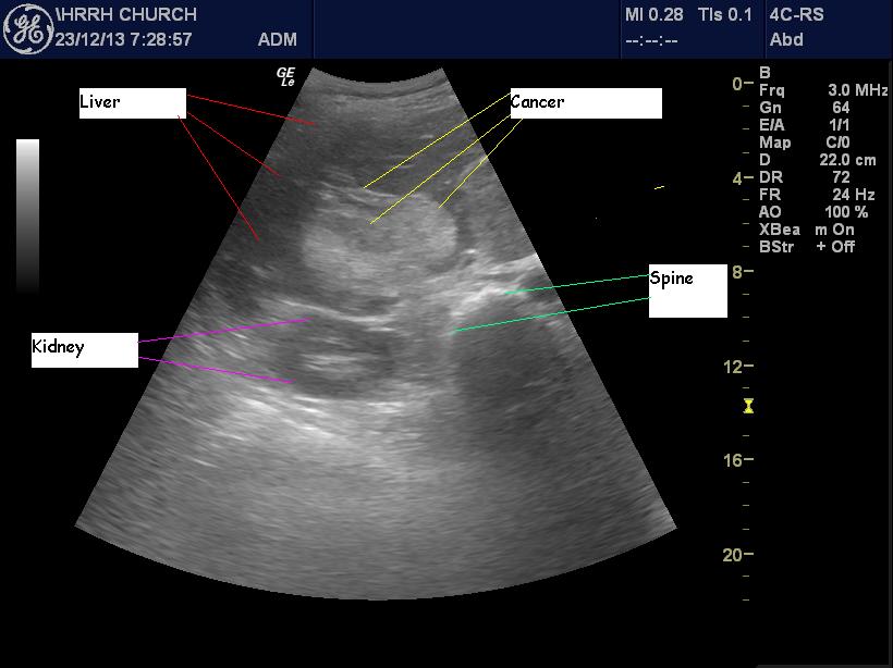
The second case was seen by Dr. Gordon:
This patient had serious COPD.
I was looking at his heart (which was Ok) and noted the huge cannonballs of cancer (previously undiagnosed) in his liver.
Every now and then the liver texture will be helpful, as here and in liver abscesses. Pretty easy to see, too.