My twinkle is better than yours

Case courtesy of Dr. Joel Turner, Fellowship Director EM Ultrasound, McGill University:
59 year old male with a previous history of renal colic presents with severe LLQ pain, and mild dysuria. He had no fever, no GI symptoms, and was a non-smoker.
His urine dipstick was positive for red blood cells. No gross hematuria. While nurse is getting appropriate pain control, I scan his left flank looking for hydronephrosis as a secondary finding of renal colic (Figure 1)
Figure 1 revealed mild hydronephrosis (and an incidental renal cyst). So, Mild hydro and Positive UA; I am done! Pain control and discharge.
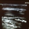
I then decide to scan his bladder (figure 2)
Low and behold: a stone is actually visualized in the left UVJ!
Finally, I throw some colour on the clip (figure 3): The greatest example of twinkling artefact you will ever see!