
Saving Brainspace with POCUS
February 8th, 2018
Here is a cool case that Lloyd Gordon recently sent us...
"A 60 year-old woman had a fever of 39.[...]


Hydronephrosis is a nice thing to see. Generally speaking you know the diagnosis when you see it. When it’s severe it’s pretty obvious. One of the pictures here is from a patient with a blocked nephrostomy tube. The pelvis is basically blown up like a balloon in the center of the kidney. Not too hard […]

It’s easy to forget that POCUS not only increases our success and reduces our complication rates for inserting central lines, it also helps us avoid putting lines where they don’t belong! While most clots will be visible a significant number can only be appreciated by the lack of compressibility of the vein. Below is another […]

This patient had LLQ pain. POCUS with the linear transducer and virtual concave on showed a tender area over the colon here and a small area of free fluid with the small bowel peristalsing around in the fluid. From this I made the diagnosis of diverticulitis. The CT confirmed this: a small area of oedematous sigmoid […]

This child was ~ 5 years old. Previously well. One day history of lots of vomiting and diarrhea. Looked pretty wiped out. Nothing specific to find on exam. POCUS revealed the maximum IVC diameter to be much less than the aortic diameter. According to some paediatric studies in normal patients the two diameters should be […]

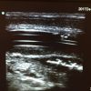
A 57 year old woman presented to the ED with left leg pain and swelling for a week. She had no past medical history, and no risk factors for DVT. On examination there was a palpable superficial cord running along the medial leg from the knee to the groin. This cord was erythematous, warm and […]

Dr. Andrea Unger is an emergency physician and avid POCUS educator. She has recorded some of the scariest images you will ever see when it comes to abscesses, reinforcing why ultrasound should be a crucial part of your exam of potential fluid collections. [Ed.] I was asked to come and see a patient, by a […]

This patient complained of vomiting, hematemesis, abdominal distension. POCUS revealed normal looking small bowel with normal peristalsis in the pelvis and lower abdomen. The upper abdomen had a few lengths of SB which were distended (SBO) and with poor peristalsis. CT confirmed small bowel obstruction plus revealed tiny areas of free air (micro-perforation). […]

Dr. Gordon is fast becoming an expert not only at performing various applications but remembering to put together great images and video for teaching others. And don’t forget, the patient’s family members can make great assistants! Below is his first submission to the EDE Blog but I have no doubt there will be many […]

I was called into an exam room by our physician assistant the other day. She had just done a POCUS on a 43 year old female, gravida 5, 10 weeks pregnant, with vaginal spotting and no significant pain. We are lucky to have some keen PA’s who are completing their certification in basic POCUS. This […]
February 8th, 2018

February 1st, 2018
January 31st, 2018
Recent Comments