
Saving Brainspace with POCUS
February 8th, 2018
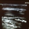
Here is a cool case that Lloyd Gordon recently sent us...
"A 60 year-old woman had a fever of 39.[...]


Diagnosing pulmonary embolism (PE) in the emergency department remains a significant challenge. Deciding on who to scan, who to anticoagulate, and who to discharge home can be difficult. This recent article by Nazerian, et al, in Chest may allow a way for POCUS to significantly rule out PE in a large number of patients. They used […]

It was 10pm on a busy shift in the emergency department. A 69 year-old man presented with sudden onset retrosternal chest pain radiating to his back. The pain lasted an hour and then resolved spontaneously. He drove himself to the ED for assessment. His initial ECG at triage was normal. While in the ED, he […]

Anton Helman (@EMCases) put up a question on Twitter regarding pregnant trauma patients and whether or not one can see a tiny amount of free fluid later in pregnancy that is physiologic. Or should one assume that it is blood. The answer is the latter. There is actually a Best BETS on using FAST in […]

Journal of Ultrasound in Medicine: Hospital-Wide Survey of Bacterial Contamination of Point-of-Care Ultrasound Probes and Coupling Gel I find the results of this article surprising but welcome. If your shop is anything like mine, it isn’t unheard of to see a probe covered in partly dried gel or even some blood in the resuscitation room. There […]

Ray and I have been teaching bedside ultrasound to the medical students at NOSM for the last couple of years. I taught them Renal/Bladder EDE last month. Kudos to Andrew Skinner from St. Paul’s in Vancouver for creating the powerpoint. I added measurement of the kidney in the long axis to the Renal part of […]

Hydronephrosis is a nice thing to see. Generally speaking you know the diagnosis when you see it. When it’s severe it’s pretty obvious. One of the pictures here is from a patient with a blocked nephrostomy tube. The pelvis is basically blown up like a balloon in the center of the kidney. Not too hard […]

Anton Helman (@EM_Cases) from EM Cases sent a tweet asking about the lung pulse and if we use and recommend it. We do. It’s mentioned at the end of the Thoracic chapter by Ben Ho. Jordan Chenkin (@POCUS_Toronto) presented Airway EDE at EDE 3 last month and spoke of the lung pulse. We will include […]

The 41st EDE 2 course is officially complete! We had a fantastic group of enthusiastic physicians attend despite the arctic temperatures. Sure there were cars that wouldn’t start and the ultrasound models’ goosebumps made it harder to scan them, but we are Canadian and we laugh at the cold! Particularly when it isn’t us getting […]

It’s easy to forget that POCUS not only increases our success and reduces our complication rates for inserting central lines, it also helps us avoid putting lines where they don’t belong! While most clots will be visible a significant number can only be appreciated by the lack of compressibility of the vein. Below is another […]

This patient had LLQ pain. POCUS with the linear transducer and virtual concave on showed a tender area over the colon here and a small area of free fluid with the small bowel peristalsing around in the fluid. From this I made the diagnosis of diverticulitis. The CT confirmed this: a small area of oedematous sigmoid […]
February 8th, 2018

February 1st, 2018
January 31st, 2018
Recent Comments