Skull fractures with #POCUS
Have you ever used POCUS to diagnose a skull fracture? We talk about it briefly at EDE 2, although we focus on the distal radius, extremities, and some other bones a bit more. But POCUS is really useful for skull fractures. Of course, if you have ready access to a CT scan, its utility will be less. But the farther you get from a CT scanner, the more you might find yourself reaching for your probe, especially in a low-risk patient, or a young patient. Here are a couple of fairly recent articles by Rabiner and Parri for the use of POCUS to diagnose skull fractures in children. And here is a case:
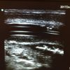
A 7 week old male was minding his own business when his playful 3-year-old brother dropped him…head-first…on a hardwood floor. The child cried right away. His (naturally) worried mom brought him to the ED. He had a fairly innocuous looking bruise to the right occiput. It was tender and the child cried whenever that area was touched. If the patient was 3 years old and had a harder skull, the management plan may have only been reassurance and that’s about it. But, at 7 weeks, the skull is relatively thin. But who wants to do a CT scan on a 7 week old? POCUS to the rescue! The bruise and surrounding area was scanned. The trouble with the infant skull is suture lines! They have an uncanny resemblance to a fracture. We all learn about suture lines in medical school, but a quick refresher of an online resource for the location of the suture lines might be in order before you check an infant for a skull fracture with POCUS. Here is this child’s scan:
There are 2 areas of the skull that look like a fracture. But the one on screen left is a normal suture line. The one on screen right is indeed a fracture. The giveaway was that each time the probe was placed directly over the apparent fracture line, the child cried. [As an aide, just like sonographic Murphy’s or sonographic McBurney’s signs, sonographic tenderness anywhere is really useful to help decide if what you are seeing on the screen is the source of the patient’s pain.]
A confirmatory elective ultrasound done in the radiology department yielded the same result. As well, their check of the intracranial contents yielded no evidence of intracranial hemorrhage. And yes, I will be checking for that myself if and when another similar case arises! The child was admitted for observation and did well.