Small Bowel Obstruction is not hard to find with POCUS: Lloyd Gordon makes his case
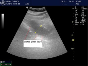
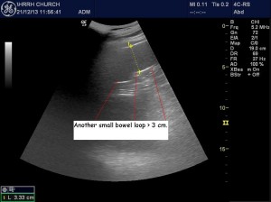
This patient complained of vomiting, hematemesis, abdominal distension. POCUS revealed normal looking small bowel with normal peristalsis in the pelvis and lower abdomen. The upper abdomen had a few lengths of SB which were distended (SBO) and with poor peristalsis. CT confirmed small bowel obstruction plus revealed tiny areas of free air (micro-perforation).
It’s really not that hard to image SB. Just take a moment when you are looking around, especially in the pelvis to get an idea of normal SB and normal peristalsis. It looks like little blobs or lengths or curves filled with granular looking fluid/contents (like some abscesses/hemorrhagic collections) that are swishing around in a characteristic way.
Then when you see abnormal bowel: distended, thickened, tender often with poor peristalsis, you will be able to suspect SBO/IBD/ischemia.


