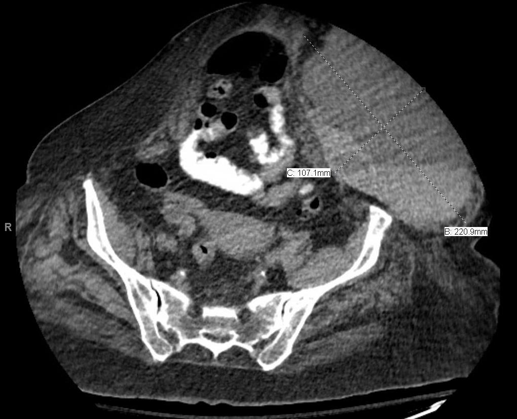
Abscesses, hematomas, and cellulitis, oh my!
I used to think that hematomas and abscesses were pretty straightforward to diagnose clinically. But I have had several cases that proved my initial suspicion to be wrong. Certainly the literature suggests we could do better differentiating cellulitis, DVT, and abscesses.
I saw a patient who presented after knee surgery with a hot, swollen and painful calf that looked exactly like a DVT. Ultrasound was not available so I followed the usual protocol of ordering low molecular weight heparin and arranging an elective the next morning to rule out DVT. Unfortunately the patient had a post-op hemorrhage into the muscle rather than a DVT, so the empiric treatment was less than ideal.
I watched a surgeon I & D what he thought was an abscess despite my POCUS showing flow. It turned out to be a tiny pseudo-aneurysm. Ouch!
Patients getting day three of their IV antibiotics in our low acuity area for cellulitis will not uncommonly demonstrate abscesses not clinically apparent. So we all have room to improve our diagnosis of clots and pus.
Lloyd Gordon has submitted a couple cases of his own:
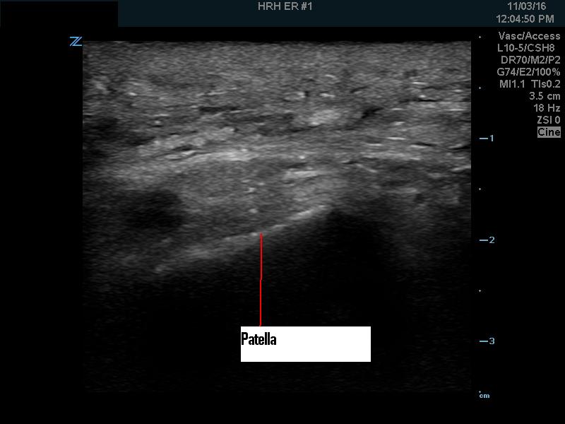
This patient was referred by a walk-in clinic because his leg looked infected. He was laying carpet earlier and had traumatized his knee. He was otherwise well. The leg had become red, swollen and painful to the point where he couldn’t walk. There was no fever.
The entire area around the knee was very boggy, red, indurated and tender. What to do? I suspect the reflex now would be to order the “cure-all”, IV antibiotics and leave it to the next person when it didn’t get better.
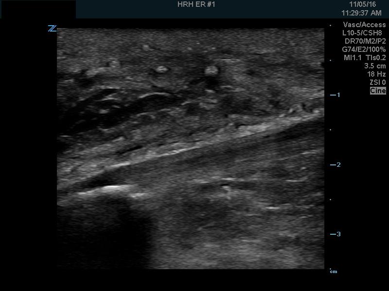
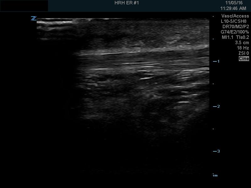
A minute or two of POCUS revealed generalized soft tissue edema from about 1/3 up from the knee to 1/3 down from the knee. There was absolutely no fluid in the knee joint so a septic knee joint seemed unlikely.
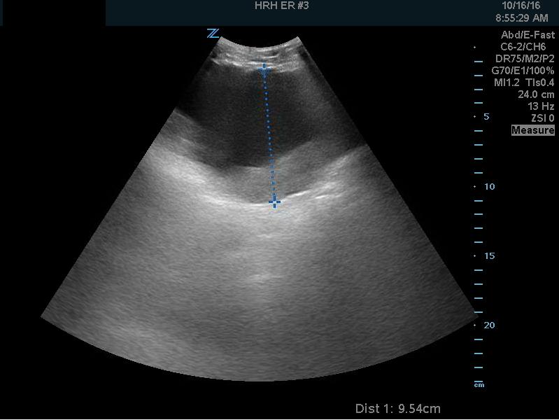
Here’s another similar case.
So avoid leaving these to your colleagues (who may leave them to their colleagues……..). POCUS the infected collection and drain it.


