Saving Brainspace with POCUS
February 8th, 2018

Editor’s note: Testicular torsion is a scary condition that we can’t afford to miss. It cannot be diagnosed on history and physical alone in almost 50% of cases so ultrasound is crucial to decision-making. POCUS can be extremely helpful in detecting the torted testis but it is important to understand that a partially torted, or […]

I really don’t like doing an I&D and then finding nothing or no pus anyway. I find it helpful to know just where the pus is, where it goes, if there’s something else besides pus, or if it needs serious surgical intervention. Once I looked at neck swelling that didn’t look too horrible but the […]

With POCUS we teach beginners to focus on the most basic knobology, physics, and imaging of the area that will answer their simple clinical question. When mastering the FAST scan it’s all about focussing on the free fluid, don’t get distracted by anything else going on. With more experience however, we all start to appreciate […]

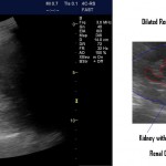
Hydronephrosis is a nice thing to see. Generally speaking you know the diagnosis when you see it. When it’s severe it’s pretty obvious. One of the pictures here is from a patient with a blocked nephrostomy tube. The pelvis is basically blown up like a balloon in the center of the kidney. Not too hard […]

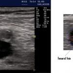
It’s easy to forget that POCUS not only increases our success and reduces our complication rates for inserting central lines, it also helps us avoid putting lines where they don’t belong! While most clots will be visible a significant number can only be appreciated by the lack of compressibility of the vein. Below is another […]

This patient had LLQ pain. POCUS with the linear transducer and virtual concave on showed a tender area over the colon here and a small area of free fluid with the small bowel peristalsing around in the fluid. From this I made the diagnosis of diverticulitis. The CT confirmed this: a small area of oedematous sigmoid […]

This child was ~ 5 years old. Previously well. One day history of lots of vomiting and diarrhea. Looked pretty wiped out. Nothing specific to find on exam. POCUS revealed the maximum IVC diameter to be much less than the aortic diameter. According to some paediatric studies in normal patients the two diameters should be […]

This patient complained of vomiting, hematemesis, abdominal distension. POCUS revealed normal looking small bowel with normal peristalsis in the pelvis and lower abdomen. The upper abdomen had a few lengths of SB which were distended (SBO) and with poor peristalsis. CT confirmed small bowel obstruction plus revealed tiny areas of free air (micro-perforation). […]

Dr. Gordon is fast becoming an expert not only at performing various applications but remembering to put together great images and video for teaching others. And don’t forget, the patient’s family members can make great assistants! Below is his first submission to the EDE Blog but I have no doubt there will be many […]
Recent Comments