Saving Brainspace with POCUS
February 8th, 2018

Here is a cool case that Lloyd Gordon recently sent us… “A 60 year-old woman had a fever of 39.6C and vomiting. The triage note mentioned abdominal pain but she didn’t have any pain when I saw her and she never asked for analgesics. Her abdomen was completely benign and she looked well. Not much […]

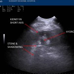
Case courtesy of Dr. Joel Turner, Fellowship Director EM Ultrasound, McGill University: 59 year old male with a previous history of renal colic presents with severe LLQ pain, and mild dysuria. He had no fever, no GI symptoms, and was a non-smoker. His urine dipstick was positive for red blood cells. No gross hematuria. While […]

Dr. Gordon shares the findings from just three recent ED shifts. There are many negative and indeterminate scans here but it provides a glimpse into how POCUS is included in the thought process for risk stratification and clinical decision making. This patient complained of R flank pain. POCUS revealed a normal kidney, uterus and pelvis. […]

We see LOTS of kidney stones in Sudbury. I’d swear that they mostly contain nickel and not calcium! About 5 years ago, I saw a 31-year-old patient with renal colic. I looked up their medical records on the computer. They had had 12 (!) CT scans in the previous 2 years. Can we do better? […]

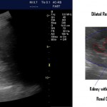
Hydronephrosis is a nice thing to see. Generally speaking you know the diagnosis when you see it. When it’s severe it’s pretty obvious. One of the pictures here is from a patient with a blocked nephrostomy tube. The pelvis is basically blown up like a balloon in the center of the kidney. Not too hard […]
Recent Comments