
Saving Brainspace with POCUS
February 8th, 2018
Here is a cool case that Lloyd Gordon recently sent us...
"A 60 year-old woman had a fever of 39.[...]


Dr. Joel Turner , the fellowship director in Emergency Medicine Ultrasound in McGill’s department of Emergency Medicine at the Jewish General Hospital in Montreal presents the following case. If this doesn’t make you reach for your ultrasound probe, nothing will! 55 year old female sent to the ED because of progressive, non-traumatic swelling of the […]

Greg posted a couple of cool cases of necrotizing fasciitis in December. We had a case of nec fasc in our department in the last few months that has made everyone quite wary! It has dropped our threshold for worrying about this nasty entity. I saw a patient last month who presented with diffuse cellulitis […]

Editors note: Lloyd has provided several great biliary scans to the blog and in his previous CBD related post talked about how an incidental finding of dilated CBD helped guide him towards diagnosing a patient with pancreatic cancer. While all of us at the EDE blog are POCUS enthusiasts, we must acknowledge that there is […]

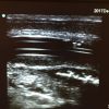
This asymptomatic 7 month old had fallen on New Year’s and the parents noted a dent in the head. There did seem to be a contusion in the left fronto-parietal area with maybe an associated “dent”. The child was normally squirmy (and very happy) so the area of interest would swim in and out of […]

Thoracic EDE is one of the hot topics in the POCUS world. This is Ben Ho’s chapter at EDE 2. Joel Turner (@JTMcGillEM) has presented some newer aspects at EDE 3. Here is a case from Dr Bernard Richard in Valleyfield that illustrates its utility. Incidentally , Bernard will be making his debut as an […]

I was looking at a patient’s kidney. There was clear hydronephrosis and proximal hydroureter. The picture I included isn’t that great because I was trying to get a good picture of the proximal ureteric stone (~ 7 mm on KUB). I was looking at the bladder for a […]

Warning: Scary article ahead. Point-of-Care Ultrasound Diagnosis of Necrotizing Fasciitis Missed By Computed Tomography and Magnetic Resonance Imaging It’s one of those diagnoses that scares us. Hard to figure out clinically in many cases at initial presentation. Adjunctive tests can be false negative. How can we increase our sensitivity? Use your POCUS skills of […]

This patient who was known to be pregnant came in quite shocky. POCUS showed blood around the spleen only, none in Morison’s Pouch or in the pelvis. A bit weird? Maybe because the ectopic was in the left tube? The patient went right to the OR without any further imaging and did well. [Editor’s note: […]

This fellow had an airbag/steering wheel injury. His sternum was tender over the fracture seen on POCUS. These are amazingly easy to find. Using a linear probe in the longitudinal plane, slide it cephalad and caudad along the sternum. I doubt this would have been seen on X-ray, the displacement is around 1 mm. [Editor’s […]

This is from EDE2. You use the linear probe and slide up the lateral radius up to the snuff box looking for a sort of saddle back mountain shape (on the normal side). You can see the fracture discontinuity on the injured side. [Editor’s note: POCUS is good to rule in a scaphoid fracture. But […]
February 8th, 2018

February 1st, 2018
January 31st, 2018
Recent Comments