
Saving Brainspace with POCUS
February 8th, 2018
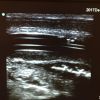
Here is a cool case that Lloyd Gordon recently sent us...
"A 60 year-old woman had a fever of 39.[...]


This patient initially gave a history of two months of upper abdominal pain. I was looking at his GB, which seemed normal when I came upon this tubular structure with no flow on Doppler. It certainly looked to be a (very dilated) CBD. Once I saw it, it was natural to ask […]

I tend to think of pericardial effusions (PCE) as being very uniform around the heart. This one was very localized to the area noted on the image. The CT Abd. (I didn’t order it!) showed the same PCE. The history was severe diarrhea and weakness. I was a bit worried about the dilated right ventricle […]

This patient had a displaced looking wrist. However the POCUS didn’t look particularly displaced at all. I was wondering about the discrepancy but not enough to get the old brain in gear (do U/S waves cause dementia in the operator?). I should have learned from my last POCUS on a Smith’s that they look very […]

Here’s a neat case from Dr Bernard Richard from Valleyfield in la belle province… A 30 y.o. man fell on a metal rod a week ago. He was seen in the ED and had his 10 cm right axillary wound sutured. I saw him a week later for 4 cm lump that was located 15 […]

I reviewed the computer charting, elderly lady with one day history of fever, vomiting, diarrhea and confusion. Sounded like a boring old gastro, give her fluids, take cultures and refer. No need to drag the U/S to the room when I’m already behind and pressed for time. Still, spare the probe and spoil the patient. […]

Scrotal EDE is one of the topics that Greg presents at EDE 3. While not a life-threatening entity (try telling that to an 18 y.o. male!), it still carries with it significant morbidity. Ultrasound is key to the diagnosis and can help assess the success of attempts at de-torsion. I recently saw an 18 y.o. […]

This patient was in his 70s with the usual litany of diseases and medications. He actually looked quite well. He complained of pain from his R neck to his R flank. The physical exam was not helpful. I had no inkling of what was wrong. I POCUS’ed his carotids/jugulars/CVS/Aorta/Kidneys/liver/spleen/bladder and all looked fine. His GB […]

This patient came in with jaundice and the LFTs were elevated. They had had a cholecystectomy in the past. POCUS revealed a large dilated fluid-filled structure in the RUQ. After looking at the kidneys/liver +/- Doppler it became clear it was a very dilated CBD. CT confirmed the biliary dilatation and obstructing mass. [Ed. note: […]

An elderly woman was sent to the ER by her senior’s residence in the middle of the night for bloody stools. The triage nurse found out that there was no blood but there was pus coming from her rectum. There was a very red and swollen area starting from the anus and extending onto her […]

A young man had clinically a distal radius fracture. The X-ray looked negative to myself and the radiologist. But with a little bit of fiddling I found the # on U/S. Before I would have said this was a Salter fracture through the epiphysis. POCUS clearly showed the # proximal to the epiphysis. Another fellow […]
February 8th, 2018

February 1st, 2018
January 31st, 2018
Recent Comments